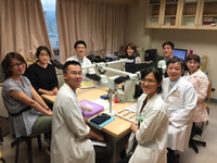
The dermatopathology laboratory has a 10-headed microscope and connected with a digital camera. Clinical images are available for every patient received skin biopsy. The lab can access clinical information and images immediately. The clinical-pathological correlation is proceeded in every case before signing out the pathology report.
The immunofluorescence images are projected into a high-resolution screen for students and trainees and recoreded by digital camera. A large immunofluorescence image archive (more than 10,000 images) is available for comparison and leraning.
There are many reference books and e-resources of journal articles. A comprehensive and well-organized slides are available for self-learning, including slides in 36 topic, more than 800 teaching slides and more than 1,600 teaching slides from self-assessment slides collected every month since 2003.
There is also a collection of previous cases of clinical-pathological conference since 2002. There are more than 2,000 slides. The database also include clinical images, powerpoint presentation file, and reference files.

